Calcaneus Fracture
Posted on 27th Jun 2017 / Published in: Ankle

A fracture of the heel bone can be disabling. Most of the time, these injuries are the result of trauma. Motor vehicle crashes and falls from elevated heights prove to be the most common reasons why this injury occurs in the first place. However, sometimes less high trauma events can also cause a fracture like jumping from a low wall and landing directly on the heel.
The calcaneus is the most fractured of all tarsal bones. Tarsal fractures account for roughly 2 percent of all fractures in adults. Out of those fractures, 60 percent are classified as a calcaneus fracture. Heel bones are often injured in a collision where other skeletal parts sustain injury as well. In up to 10 percent of those cases, the patient will also end up with a fracture in their hip, spine or other calcaneus. Injuries to this region often cause damage to the subtalar joint and cause stiffness in the joint. The injury makes it quite difficult to walk on a slanted surface or uneven ground.
Calcaneus Fracture Anatomy
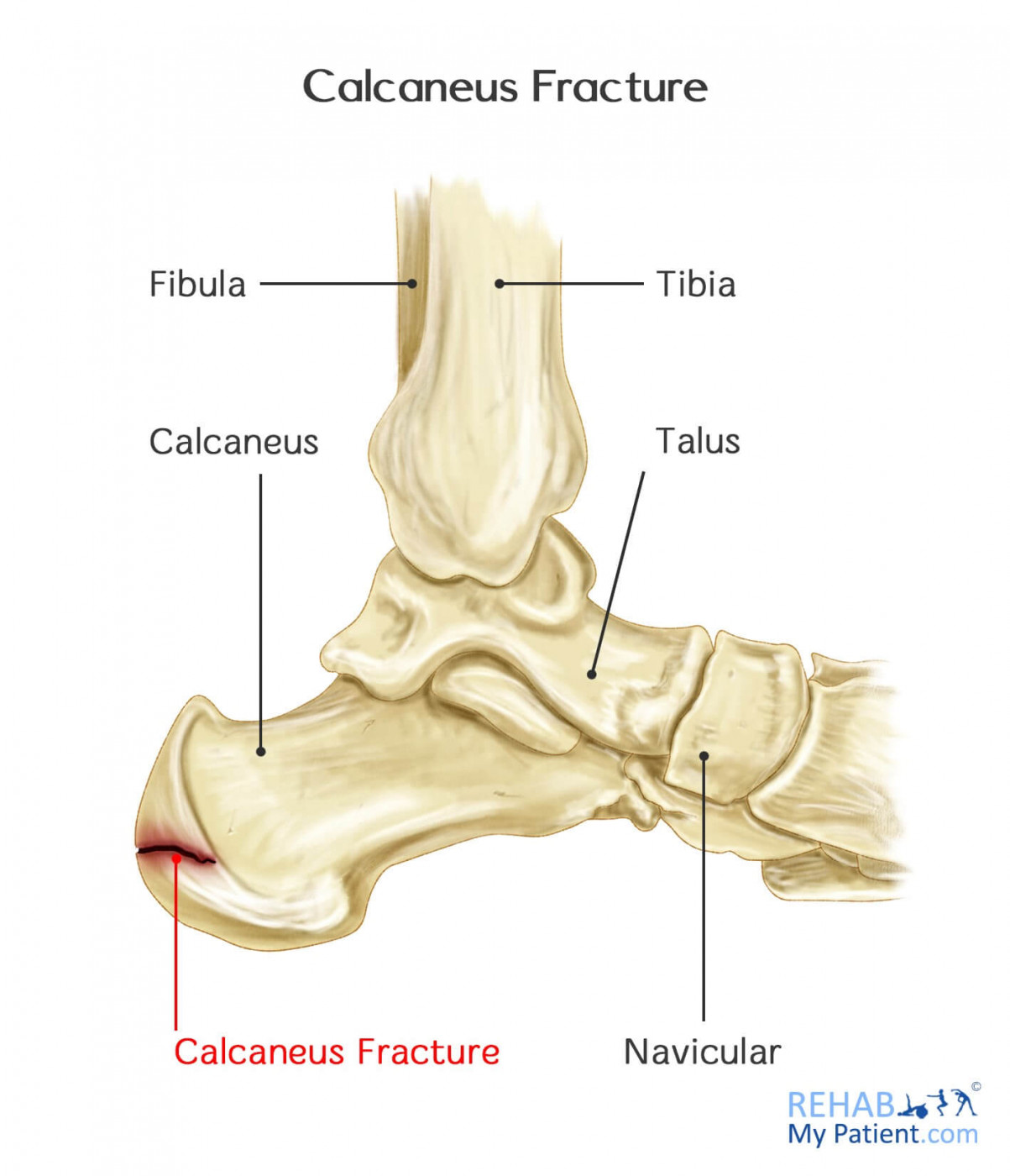
To gain a better understanding of the fracture, you need to understand the foot's anatomy. The foot is composed of bones, blood vessels, nerves, strong bands holding the bones together (ligaments), strong bands attaching the muscles to the bones (tendons), fatty tissue, muscle, skin and nails.

A small calcaneal fracture

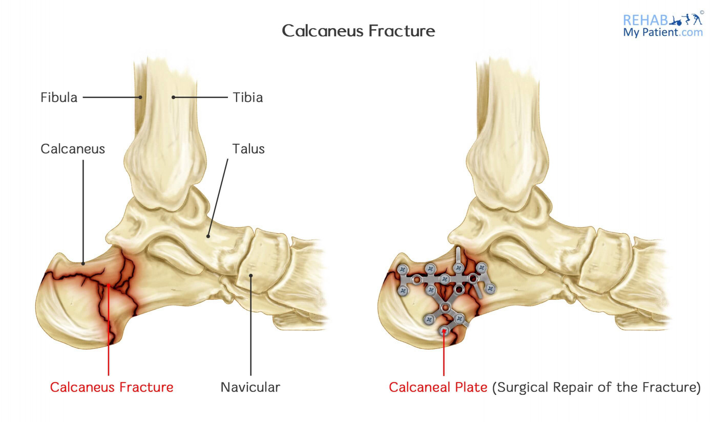
A large calcaneal fracture and the surgical operation to attach the pieces of bone
How to Treat Calcaneus Fractures:
When it comes to developing a treatment plan, a few things need to be considered, such as:
- Overall health
- Injury cause
- Injury severity
- Soft tissue damage extent
Since the majority of fractures cause the bone to widen, the treatment goal is to restore your heel's normal anatomy. Patients that have their normal anatomy restored will have a better outcome than those who don’t. Recreating the anatomy will often involve surgery, which poses an increased risk of complications.
- Nonsurgical Treatment
If there are no pieces of broken bone that were displaced from the injury, surgery might not be required. Casting and immobilization might be an option to keep the broken ends properly positioned while healing. Weight cannot be placed on the foot until the bone has a chance to heal completely, which can take six to eight weeks, if not longer.
- Surgical Treatment
Open reduction and internal fixation involves repositioning the bone fragments into normal alignment. Special screws are used to hold everything together. If the bone pieces are too large, percutaneous screw fixation can move the pieces back into place by pushing on them without requiring a large incision. Specialized screws can be put through the small incision to help keep the pieces of the bone together.
Tips:
- If you begin struggling to walk or move the foot, you need to have it examined for a calcaneus fracture.
- Puncture wounds and lacerations need to be addressed quickly to determine the cause.
- Red streaks traveling up the leg might be a sign of a fracture in the foot.
- If the toes and feet become numb, you might have bone pieces that are out of place in the foot.
- Foot pain and swelling that worsens should be address with your health care provider.
- Therapy can be useful for many reasons. Low intensity pulsed ultrasound can speed up fracture repair, and treatment can be done to the leg to reduce the effects of altered gait. Concentrating on reducing inflammation around the fracture site can also aid healing.
- If you were surprised that you sustained a fracture (i.e. the trauma was not particularly heavy), you should consider a DEXA (bone density scan) to make sure you do not have osteoporosis or bone thinning.
Sign UP
Sign up for your free trial now!
Get started with Rehab My Patient today and revolutionize your exercise prescription process for effective rehabilitation.
Start Your 14-Day Free Trial