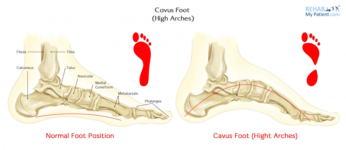
Cavus Foot (High Arches)
Posted on 09th Jul 2017 / Published in: Ankle

Cavus foot is a condition where the foot has an extremely high arch. Due to the higher arch, a tremendous amount of weight is placed onto the heel and ball of the foot when standing or walking. Cavus foot can cause any number of symptoms and signs, such as instability and pain. The condition can develop at any point in time. One or both feet can be affected.
Cavus foot, known often as pes cavus, is often just a variation of normal, so should not necessarily be seen as a pathology. In fact in the middle ages in England, it was seen as a sign of the aristocracy.
Cavus Foot Anatomy
Cavus foot is commonly used to describe a spectrum of different foot shapes that all have a high arch in common. The components of the cavus are an increased pitch and varus within the hind foot, adduction and varus within the forefoot and plantar flexion within the midfoot. The cavus shape tends to be associated with any changes within the mechanics of the foot. It is significantly less balanced and flexible than that of a neutral foot. Even though 1/5 to 1/4 of the population has the condition, many people do not have any symptoms or problems as a result of pes cavus.
How to Treat Cavus Foot:
-
Physical Therapy and Podiatry
Physical therapy and podiatry might be prescribed to help strengthen and stretch the muscles within the lower leg. Weak muscles on the outside of the lower leg and tight calf muscles tend to be present in this condition. Even though therapy might not be able to change the shape of the foot, it can help with controlling function and pain. Since the foot is often rolled inward accompanied by the high arch, the individual is prone to a chronic ankle sprain. Using reactive muscle strengthening can be beneficial, as well as ankle bracing. Your therapist might also look at your gait.
- Tendon Lengthening
This particular procedure involves making small cuts within the tight tendons of the lower leg to provide the foot with better alignment. After surgery, a period of immobilization for multiple weeks is required to let the tendons heal.
- Osteotomy
If the condition has existed since childhood and the bony structure of the foot hasn’t grown properly, small sections of the bone might need to be removed to restore proper positioning of the foot. The first metatarsal, which is located in the midfoot behind your big toe, is often treated with this procedure. Most of the time, the metatarsal is positioned at a downward angle greater than normal, which helps to roll the ankle outward when the person bears weight on the foot. This procedure helps to normalize the angle and put the foot into a neutral position. There are times when this procedure is performed in conjunction with soft tissue surgery.
- Arthrodesis
This particular procedure will lock the joint affected into a fixed position. Even though this procedure is a last resort, it sometimes proves necessary to correct the deformity or when arthritis is found.
Tips:
- If you notice you have a particularly high instep, you may have pes cavus.
- An unstable foot from the heel tilting inward could end up leading to ankle sprains.
- If calluses develop on the side, ball or heel of the feet, you need to have that checked out.
A podiatrist will be able to assess the problem.
- Hammertoes or claw toes could be a sign of cavus foot.
- Genetics plays a key role in who is afflicted with cavus foot.
Sign UP
Sign up for your free trial now!
Get started with Rehab My Patient today and revolutionize your exercise prescription process for effective rehabilitation.
Start Your 14-Day Free Trial