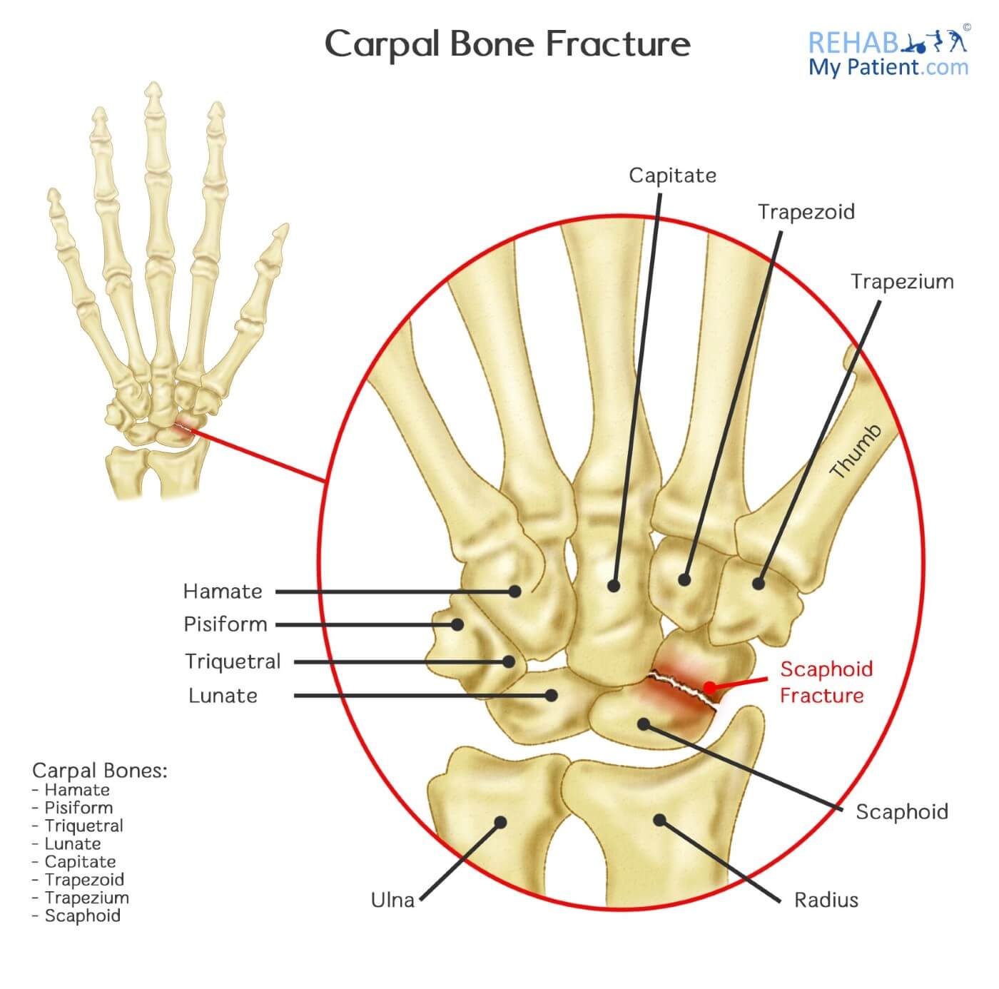
Carpal Bone Fracture
Posted on 17th Oct 2016 / Published in: Hand/Fingers/Thumb , Wrist

An individual with a wrist fracture is someone who has broken one of the 10 different bones making up the wrist joint. With 10 different bones in the wrist: eight carpal bones and two forearm bones, wrist fractures tend to be one of the most common types of fractures seen in individuals entering the emergency room.
By far the most common cause of injury occurs from a fall. However, other injuries can occur with sports injuries (e.g. hitting a tree stump during golf, trauma during contact sports, or a road traffic accident).
Carpal Bone Fracture Anatomy
Nestled between the hand and the forearm, the wrist extends from the insertion of the pronator quadrates of the ulna and the radius all the way to the carpometacarpal joints. With eight carpal bones, all of them are associated with the network of interwoven ligament connections.
The ulna and radial arteries provide the vascular supply of the wrist, as well as that of the posterior and anterior interosseous arteries. All of the vessels contribute to palmar formation and dorsal vascular arches providing circulation to that of the carpal bones. The blood flow pattern is of importance. The scaphoid gets its blood supply from two of the small branches primarily arising from the radial artery. Circulation to that of the proximal pole is maintained from the intraosseous vessels.

Due to the vascular supply for the pole relying upon the vessels that enter the scaphoid, more injuries to the wrist cause a disruption in blood flow. Fractures of the wrist can interfere with proximal flow, which places the scaphoid at risk for developing avascular necrosis.
What to Do if You Think You Have Fractured Your Wrist
First, go to hospital and ask the doctor for a physical examination. They will check the wrist and advise if an X-ray is required. If you cannot get to hospital, seeing a physical therapist or your General Practitioner can also be useful to diagnose a wrist fracture. If you have swelling around your wrist, gross stiffness, and cannot put any weight through the wrist (as if you were leaning your palm flat on a table), then you might have a wrist fracture.
How to Treat Carpal Bone Fractures:
- Immobilization of the Wrist
Treatment of a fracture will often consist of immobilization of the wrist using a cast that extends from the forearm all the way to the knuckles, which leaves the fingers and thumb free to move. In the event the scaphoid bone is involved in the fracture, the cast will extend beyond the elbow for the first six weeks, as well as incorporating the thumb into the cast. X-rays will be required to follow-up on the healing and determine if there is any loss of the reduction. Wrists can be immobilized for three to four months to complete the healing process.
- Surgery
When the bones have moved out of alignment or don’t stay aligned, surgery may be required to realign the bones, restore function and ensure the area heals properly. Surgery is often an open procedure, but there are surgeons who prefer to reduce the fracture arthroscopically, using a small incision and a miniscule camera and lens. Surgery might involve internal or external fixation with wires, pins, plates and screws, especially when the distal radius bone is fractured. Hardware can be removed later on after recovered.
- Therapy
Later stage treatment and therapy can aid significantly following wrist or carpal bone fracture. Once a cast is removed, the wrist will likely be very stiff. Therapy will mobilize the joints to get your wrist to its full range of movement. Treatment may also reduce inflammation in the wrist, and offload pressure on the surrounding forearm muscles and tendons.
Tips:
- Falls are the biggest cause of wrist fracture, take care on slippery surfaces (wear appropriate footwear, walk on the grassy part of a pavement if it’s icy, take extra care in cold, snowy or icy conditions.
- Contact sports increase your risk of fracturing your carpal bone.
- Elderly individuals are more prone to falls that could fracture the wrist.
- If your injury was quite innocuous, consider requesting a DEXA (bone density scan) from your doctor to check you are not suffering with bone thinning or osteoporosis.
- Postmenopausal women are susceptible to fracturing their wrist.
Sign UP
Sign up for your free trial now!
Get started with Rehab My Patient today and revolutionize your exercise prescription process for effective rehabilitation.
Start Your 14-Day Free Trial