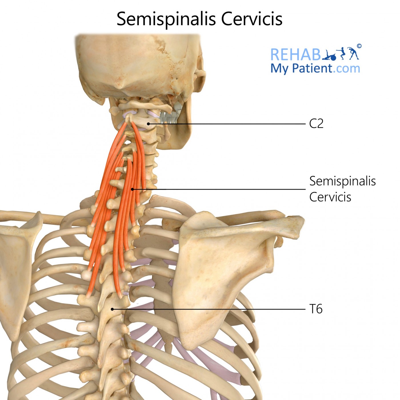
Semispinalis Cervicis
Posted on 29th Jul 2020 / Published in: Neck

General information
This muscle arises out of a series of tendinous and fleshy fibres out of the transverse processes for the upper five to six vertebrae. It is inserted into the cervical spinal processes out of the axis and to the fifth inclusive. The fasciculus that is connected to the axis is the largest and most muscular in structure. This muscle is thicker in nature than that of the Semispinalis dorsi.
Literal meaning
Half thorn of the neck.
Interesting information
Of all cervical injuries in athletes, the most common by far is that of an acute sprain or strain to the musculature in the neck, as well as any contusions to the soft tissues. Strains are an injury to the muscle when the tendon unit becomes stretched or overloaded. The cervical muscles tend to be strained more often by those who are active sports enthusiasts. Sprains mean that the structure the connects the cervical joints to the vertebrae have sustained damage. Rugby and American football players are prone to injuries here due to all of the stretching to make tackles, catch balls and throw the ball to another player.
Origin
Transverse processes of T1-T6.
Insertion
Sides of the spinous processes for C2-C5.
Function
Extension of the cervical vertebral column for rotating it to the other side.
Nerve supply
Dorsal rami of the thoracic and cervical spinal nerves C3-T5.
Blood supply
Lateral cervical muscle portion receives blood from the deep cervical artery arising from the costocervical trunk out of the subclavian artery as well as the descending branch for the occipital artery, which forms with the deep cervical artery. Dorsal branches in the posterior intercostals arteries arising out of the thoracic aorta.

Relevant research
Clinically examining the cervical spine is one of the main requirements for managing neck pain on the therapeutic and exploratory levels. This type of exam is far from becoming standard and will vary based on the training level of the individual. Osteopaths, physicians and chiropractors are all going to use different techniques and maneuvers to measure the different data. Literature has not been able to document these maneuvers consistently, which is why studies are quite rare on the muscle.
The purpose of the study was to assess the interexaminer agreement when dealing with a manual, structured and clinical examination of the neck muscles. To help correlate the data, scores in a functional questionnaire were assessed. The results showed that the interexaminer agreement was moderate and the number of tender spots found was strongly correlated with the functional impairment suffered by patients. Pain at the lower attachment point of the levator scapulae was associated with dysfunction of the median or upper cervical spine.
Maigne JY, Chantelot F, Chatellier G. Interexaminer agreement of clinical examination of the neck in manual medicine. Ann Phys Rehabil Med. 2009;52(1):41?48. doi:10.1016/j.rehab.2008.11.001
Semispinalis cervicis exercises
Chin to chest stretch
Get into a seated position on the ground. Place both the hands at the base of the head with fingers interlocked. Point the thumbs down and the elbows straight ahead. Pull the head down to the chest and hold for 20 to 30 seconds. Perform 10 repetitions of this exercise.
Sign UP
Sign up for your free trial now!
Get started with Rehab My Patient today and revolutionize your exercise prescription process for effective rehabilitation.
Start Your 14-Day Free Trial